
Análise Morfofisiológica do Osso Keratobranquial II localizado no Hyoide of the Green Turtle (Chelonias mydas) encontrado no Peruíbe, Litoral Sul do Brasil, Mosaico de Unidades de Conservação-Juréia- Itatins e APACIP – Área de Protecção Ambiental-Cananéia-Iguápe- Peruíbe-SP
ABSTRACT
The green turtle (Chelonia mydas) present in tropical seas, uses as a feeding area the
coastal region of Peruíbe, has the skull as a relatively large and solid structure, and a
strong jaw formed by the junction of small bones as it has very abrasive feeding. By
applying scanning electron microscopy techniques, it was possible to identify the
presence of a bone structure located in the hyoid in the ventral region of the skull along
with the mandible of juvenile individuals of green turtles, and as there is no related
research, it was necessary to perform a CT scan, decalcification and histology of the
quelonian hyoid, to discover the morphological composition of this new structure,
described only in the species Chelonia mydas. Thus, the morphology of the structures and
its confirmation as a real bone, with characteristic of spongy bone, described as
certobranchial II, was confirmed, thus helping researchers to seek other ways to
understand the feeding processes of these animals that are going through a series of
serious environmental problems and therefore perhaps having to change their eating
habits to overcome the high level of pollution that we are finding in the oceans.
Keywords: anatomy, histology, morphology, tomography.
Descrição osso ceratobranquiall tartaruga verde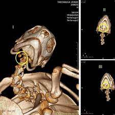
https://ibimm.org.br/analise-morfofisiologica-do-osso-keratobranquial-ii-localizado-no-hyoide-of-the-green-turtle-chelonias-mydas-encontrado-no-peruibe-litoral-sul-do-brasil-mosaico-de-unidades-de-conservacao-jureia/PRODUÇÕES CIENTÍFICAS IBIMMÚLTIMAS NOTÍCIAS !!!ABSTRACT The green turtle (Chelonia mydas) present in tropical seas, uses as a feeding area the coastal region of Peruíbe, has the skull as a relatively large and solid structure, and a strong jaw formed by the junction of small bones as it has very abrasive feeding. By applying scanning electron microscopy techniques,...IBIMMIBIMM INSTITUTO DE BIOLOGIA MARINHA E MEIO AMBIENTEcontatobiologia@ibimm.org.brEditorIBIMM - INSTITUTO BIOLOGIA MARINHA E MEIO AMBIENTE
